
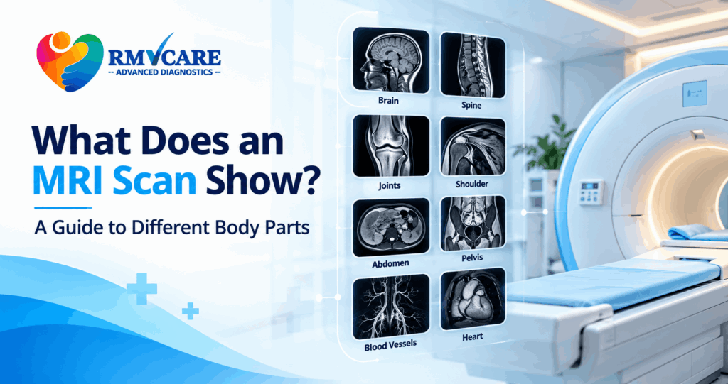
What Does an MRI Scan Show? A Guide to Different Body Parts
An MRI scan is an advanced imaging test that creates detailed images of structures inside the body using magnetic fields and radio waves. But what does an MRI scan show exactly? The answer depends on the body part being examined and the type of MRI prescribed by your doctor.
An MRI can provide detailed views of the brain, spine, joints, muscles, organs, blood vessels and other soft tissues. Unlike X-rays and CT scans, MRI does not use ionising radiation. This makes MRI particularly useful when doctors need detailed information about soft tissues and certain internal structures.
In this guide, we explain what an MRI scan can show in different parts of the body, when doctors may recommend it, and what you should know before undergoing an MRI scan.
What Is an MRI Scan?
MRI stands for Magnetic Resonance Imaging. It uses a strong magnetic field, radio waves and computer technology to create detailed images of the inside of the body.
Depending on the area being examined, an MRI scan may help doctors evaluate the brain, spine, joints, muscles, organs, blood vessels and other soft tissues.
Different MRI protocols can be used depending on the clinical question. For example, a doctor may recommend an MRI brain scan for neurological symptoms, an MRI spine for back or neck problems, or an MRI joint scan for suspected ligament or cartilage injuries.
The images are interpreted by a qualified radiologist, who prepares a report for the treating doctor.
What Does an MRI Scan Show?
An MRI can show many types of tissues and abnormalities. The exact findings depend on the body part scanned and the MRI protocol used.
Here are some of the common areas that can be examined with MRI.
1. What Does a Brain MRI Show?
A brain MRI scan provides detailed images of the brain and surrounding structures. Doctors may recommend it when they need more information about neurological symptoms or abnormalities.
A brain MRI may help evaluate:
- Brain tissue
- Certain tumours or masses
- Areas of inflammation
- Structural abnormalities
- Certain blood vessel abnormalities
- Changes associated with some neurological conditions
- Areas of abnormal tissue or fluid
Depending on the clinical requirement, specialised MRI techniques such as diffusion imaging, contrast-enhanced MRI or MR angiography may be recommended.
The choice of MRI technique depends on the symptoms and the question your doctor needs the scan to answer.
2. What Does a Spine MRI Show?
An MRI spine scan provides detailed images of the vertebrae, intervertebral discs, spinal cord, nerves and surrounding soft tissues.
Doctors may recommend an MRI of the cervical, dorsal or lumbar spine depending on the location of symptoms.
A spine MRI may help evaluate:
- Disc bulges or herniation
- Degenerative changes
- Nerve compression
- Spinal cord abnormalities
- Inflammation
- Certain infections
- Tumours or other abnormal growths
- Soft-tissue and ligament-related problems
For people experiencing persistent back pain, neck pain, numbness, tingling or weakness, a doctor may recommend MRI when detailed imaging of the spine and surrounding structures is clinically appropriate.
3. What Does a Knee MRI Show?
A knee MRI provides detailed images of the soft tissues and structures within the knee joint.
It can help doctors evaluate structures such as:
- Menisci
- Ligaments
- Tendons
- Cartilage
- Muscles
- Bone marrow
- Joint structures
A knee MRI may be recommended when there is suspected ligament injury, meniscal injury, cartilage damage or another condition that requires detailed imaging.
MRI is particularly useful for soft-tissue evaluation, which is one reason it is commonly used for musculoskeletal imaging.
4. What Does a Shoulder MRI Show?
A shoulder MRI can provide detailed images of the shoulder joint and surrounding soft tissues.
It may help evaluate:
- Rotator cuff tendons
- Labrum
- Muscles
- Tendons
- Ligaments
- Cartilage
- Joint structures
- Certain soft-tissue abnormalities
Doctors may recommend an MRI when shoulder pain, weakness, restricted movement or an injury requires detailed evaluation.
5. What Does an MRI of the Abdomen Show?
An abdominal MRI can provide detailed images of several organs and structures within the abdomen.
Depending on the examination, MRI may be used to assess:
- Liver
- Pancreas
- Kidneys
- Spleen
- Biliary system
- Adrenal glands
- Blood vessels
- Other abdominal soft tissues
Specialised examinations such as MRCP can provide detailed imaging of the bile ducts and pancreatic duct.
An abdominal MRI may be recommended when a doctor needs additional information about a suspected abnormality or when MRI provides useful soft-tissue characterisation.
6. What Does a Pelvic MRI Show?
A pelvic MRI can provide detailed images of pelvic organs and surrounding soft tissues.
Depending on the patient and clinical indication, it may be used to evaluate structures such as:
- Uterus
- Ovaries
- Urinary bladder
- Prostate
- Pelvic muscles and soft tissues
- Other pelvic structures
Doctors may recommend pelvic MRI for further evaluation of certain abnormalities identified through other examinations or when detailed soft-tissue imaging is required.
7. What Does an MRI Show in Muscles and Joints?
MRI is widely used for musculoskeletal imaging because it can provide detailed images of soft tissues.
Depending on the body part, MRI can help assess:
- Muscles
- Tendons
- Ligaments
- Cartilage
- Joints
- Bone marrow
- Soft-tissue abnormalities
MRI may therefore be used for areas such as the knee, shoulder, hip, ankle, wrist, elbow and other joints.
8. What Does an MRI Show in Blood Vessels?
Specialised MRI techniques called MR angiography (MRA) and MR venography (MRV) can be used to produce images of blood vessels.
Depending on the examination, these techniques may help doctors assess blood vessels in areas such as the:
- Brain
- Neck
- Abdomen
- Pelvis
- Extremities
The specific MRI technique depends on the clinical requirement and the vessels that need to be evaluated.
9. Can an MRI Show Tumours?
MRI can be useful in evaluating certain tumours and other abnormal masses because it provides detailed images of soft tissues.
Depending on the body part and type of examination, MRI may help doctors understand:
- The location of an abnormal mass
- Its relationship with nearby structures
- Characteristics of the surrounding tissue
- Changes over time
In some cases, a contrast-enhanced MRI may be recommended to provide additional information.
However, an MRI image alone does not necessarily establish a final diagnosis. Doctors may consider symptoms, medical history, laboratory tests, previous imaging and, when necessary, biopsy or other investigations.
MRI With Contrast vs MRI Without Contrast
Not every MRI scan requires contrast.
A contrast-enhanced MRI involves administering a contrast agent, usually through a vein, when the additional information may be clinically useful.
Contrast may help improve the visibility or characterisation of certain tissues and abnormalities.
Whether contrast is required depends on:
- The body part being examined
- The suspected condition
- The purpose of the examination
- Previous imaging findings
- Your medical history
Your doctor or radiologist will determine whether contrast is appropriate for your examination.
How Long Does an MRI Scan Take?
The duration of an MRI depends on the body part being examined, the imaging protocol and whether additional sequences or contrast are required.
An MRI examination can commonly take anywhere from around 15 to 90 minutes, depending on the examination.
During the scan, it is important to remain as still as possible so that clear images can be obtained.
Is an MRI Scan Safe?
MRI does not use ionising radiation and is considered a widely used medical imaging technique.
However, because MRI uses a strong magnetic field, patients must inform the imaging centre about certain implants, devices or metal objects in the body before the examination.
Before an MRI, tell your healthcare team if you have:
- A pacemaker or implanted medical device
- Certain metal implants
- Previous surgery involving metal implants
- Metal fragments in the body
- A history of severe claustrophobia
- Pregnancy or possible pregnancy
- Any other relevant medical condition
The MRI team will determine whether the examination is appropriate and what precautions are required.
How Should You Prepare for an MRI Scan?
MRI preparation varies according to the type of examination.
Depending on the scan, you may be asked to:
- Remove jewellery and metal accessories
- Change into suitable clothing or a gown
- Follow fasting instructions if required
- Bring previous imaging reports
- Inform the radiology team about implants or medical devices
- Follow specific instructions if contrast is planned
Always follow the preparation instructions provided by your doctor or diagnostic centre.
When Should You Get an MRI Scan?
An MRI should be performed when recommended by a qualified healthcare professional based on your symptoms, examination and medical history.
Doctors may consider MRI when they need detailed information about soft tissues, the brain, spine, joints, organs or blood vessels.
For example, depending on the clinical situation, MRI may be considered for:
- Persistent back or neck pain
- Certain neurological symptoms
- Suspected ligament or cartilage injuries
- Joint pain following injury
- Certain unexplained soft-tissue abnormalities
- Further evaluation of findings seen on another imaging test
The appropriate imaging test depends on the individual clinical situation. MRI is not automatically the right test for every symptom.
MRI Scan vs CT Scan: Which One Is Better?
MRI and CT scans are different imaging techniques and are used for different clinical purposes.
MRI uses magnetic fields and radio waves, while CT uses X-rays. MRI is particularly useful for detailed soft-tissue imaging, whereas CT can be highly useful for evaluating bones, certain chest and abdominal conditions, trauma and other situations where rapid imaging is required.
Therefore, there is no single scan that is “better” for every condition. The doctor selects the appropriate examination based on the patient’s symptoms and the information required.
What Happens After an MRI Scan?
After the MRI examination, the acquired images are reviewed by a radiologist.
The radiologist evaluates the images and prepares a diagnostic report. Your treating doctor can then interpret the findings alongside your symptoms, medical history and other test results.
Depending on the findings, your doctor may recommend monitoring, medication, additional investigations, specialist consultation or another appropriate treatment plan.
Frequently Asked Questions About MRI Scans
1. What does an MRI scan show?
An MRI scan can provide detailed images of soft tissues and various internal structures, including the brain, spine, joints, muscles, organs and blood vessels, depending on the type of MRI performed.
2. What can an MRI detect?
MRI can help doctors evaluate many abnormalities involving the brain, spine, joints, soft tissues and internal organs. The findings depend on the body part and MRI protocol.
3. Does an MRI scan use radiation?
No. MRI uses magnetic fields and radio waves rather than ionising radiation.
4. Can an MRI detect a tumour?
MRI can help identify and characterise certain masses or tumours and assess their location and relationship with surrounding structures. Additional tests may be required for a definitive diagnosis.
5. How long does an MRI scan take?
The duration varies depending on the body part and examination protocol. Many MRI examinations take approximately 15 to 90 minutes.
6. Is MRI painful?
MRI itself is generally non-invasive and does not cause pain. However, you need to remain still during the examination, and some people may feel uncomfortable inside the scanner.
7. Can I have an MRI if I have a metal implant?
Some implants and devices are compatible with MRI, while others may not be. Always inform the MRI team about any implant, device or metal fragment before your scan.
8. Does every MRI require contrast?
No. Many MRI examinations are performed without contrast. Contrast is used only when clinically indicated.
Get an MRI Scan in Kandivali, Mumbai
If your doctor has recommended an MRI scan in Kandivali, Mumbai, choosing a diagnostic centre with appropriate imaging technology and qualified radiology expertise is important.
At RM VCare Advanced Diagnostics, patients can access advanced diagnostic imaging services in Kandivali, Mumbai. The appropriate MRI examination is selected according to the doctor’s prescription and the patient’s clinical requirements.
If you have been advised to undergo an MRI scan, bring your prescription, previous reports and relevant medical information to your appointment.
RM VCare Advanced Diagnostics
Kandivali, Mumbai
Website: rmvcarediagnostics.com
Medical Disclaimer: This article is intended for general educational purposes and should not be considered a substitute for professional medical advice, diagnosis or treatment. The type of imaging test required depends on the individual’s symptoms and clinical condition. Always follow the advice of your qualified healthcare professional.