
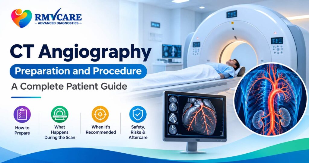
CT Angiography Preparation and Procedure: Everything You Need to Know
When doctors need detailed images of blood vessels to investigate circulation-related conditions, they may recommend a specialised imaging examination called CT angiography (CTA). This advanced diagnostic technique combines computed tomography (CT) scanning with an iodine-based contrast material to produce detailed images of blood vessels and surrounding structures.
Understanding CT angiography preparation and procedure can help patients feel more confident before their examination. Knowing what happens before, during and after the scan can also help you follow the correct instructions and avoid unnecessary delays.
Whether your doctor has recommended a CT angiogram to examine blood vessels in the brain, heart, chest, abdomen or legs, this guide explains its purpose, preparation, procedure, benefits and important safety considerations.
What Is CT Angiography?
CT angiography, also known as a CT angiogram, is a specialised diagnostic imaging examination used to visualise blood vessels throughout the body.
It uses X-rays, computer processing and an iodine-based contrast agent, usually injected into a vein, to create detailed images of arteries and selected vascular structures.
Unlike conventional CT examinations that primarily focus on organs and tissues, CT angiography is specifically designed to evaluate blood vessels and identify certain abnormalities affecting circulation.
Doctors may recommend this examination to investigate:
- Narrowed or blocked blood vessels.
- Aneurysms (abnormal widening of blood vessels).
- Certain blood clots.
- Abnormal blood vessel structures.
- Selected vascular injuries.
- Conditions affecting blood supply to organs.
The examination helps doctors understand the location and extent of certain vascular abnormalities and plan appropriate medical management.
How Does CT Angiography Work?
CT angiography works by combining CT imaging technology with intravenous contrast material.
The contrast agent travels through the bloodstream and makes blood vessels more visible on the CT images. The scanner captures multiple images as the contrast moves through the targeted blood vessels.
These images are then processed by specialised software to create detailed cross-sectional and, when appropriate, three-dimensional views.
The radiologist can examine the images to assess the structure of blood vessels, identify possible narrowing or abnormalities and evaluate surrounding anatomical structures.
What Are the Different Types of CT Angiography?
CT angiography can examine blood vessels in different parts of the body. The specific examination depends on the doctor’s recommendation and the suspected medical condition.
1. CT Brain Angiography
CT angiography of the brain examines blood vessels supplying the brain.
It may be recommended to investigate certain aneurysms, vascular abnormalities, narrowed arteries and selected conditions affecting cerebral circulation.
2. CT Coronary Angiography
CT coronary angiography, also called CCTA, examines the coronary arteries that supply blood to the heart.
It may help doctors assess coronary artery plaque and narrowing in appropriately selected patients.
3. CT Pulmonary Angiography
CT pulmonary angiography examines the blood vessels in the lungs.
It is commonly used when doctors need to investigate a suspected pulmonary embolism, a blood clot affecting the pulmonary arteries.
4. CT Aortic Angiography
This examination evaluates the aorta, the body’s largest artery.
It may be recommended to investigate conditions such as aortic aneurysms, dissections and selected vascular abnormalities.
5. CT Peripheral Angiography
CT peripheral angiography examines blood vessels in areas such as the legs and arms.
It may help assess narrowing, blockages and other vascular abnormalities that affect blood circulation in the limbs.
Why Do Doctors Recommend a CT Angiography?
Doctors may recommend CT angiography when detailed information about blood vessels is required for diagnosis or treatment planning.
Some common clinical applications include:
- Detecting aneurysms: Helps identify abnormal widening of certain blood vessels.
- Investigating blocked arteries: Helps evaluate narrowing or obstruction in selected blood vessels.
- Assessing blood clots: Can help identify certain clots, including pulmonary emboli.
- Evaluating vascular injuries: Provides detailed images of selected blood vessel injuries.
- Examining coronary arteries: Helps assess coronary artery disease in suitable patients.
- Investigating vascular abnormalities: Helps assess certain structural abnormalities in arteries and other blood vessels.
The treating doctor determines whether CT angiography is appropriate based on symptoms, clinical history and other examination findings.
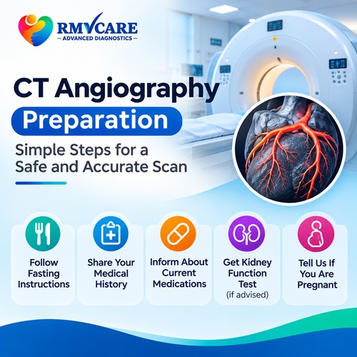
CT Angiography Preparation: What Should You Do Before the Scan?
Proper preparation is important for obtaining good-quality CT angiography images and ensuring patient safety.
Preparation instructions may vary depending on the type of angiography, the contrast material used and your medical history.
1. Inform Your Doctor About Your Medical History
Before the examination, inform your doctor and radiology team if you have:
- Kidney disease or reduced kidney function.
- A previous allergic-like reaction to CT contrast material.
- Diabetes or other significant medical conditions.
- Thyroid-related conditions, if relevant.
- A history of heart disease.
- A possibility of pregnancy.
- Any other known allergies or previous contrast-related complications.
This information helps the medical team assess potential risks and plan the appropriate examination.
2. Follow Fasting Instructions
Some CT angiography examinations require patients to avoid food or drinks for a specified period before the scan.
The fasting duration can differ according to the examination and centre-specific protocol.
Do not assume that every CT angiogram has the same fasting requirements. Confirm the exact instructions when scheduling your appointment.
3. Inform the Team About Your Medications
Provide details of all medicines you currently take, including diabetes medication and supplements.
Do not stop or change prescribed medicines without instructions from your treating doctor or radiology team.
Certain examinations, particularly coronary CT angiography, may involve additional medication instructions depending on the patient’s heart rate and clinical condition.
4. Kidney Function Assessment
Because CT angiography commonly involves iodine-based intravenous contrast, your doctor may request kidney function tests, such as serum creatinine or estimated glomerular filtration rate (eGFR).
This is particularly important for patients with known kidney disease, acute kidney injury or other relevant risk factors.
The radiology team will decide whether additional assessment or precautions are necessary.
5. Wear Comfortable Clothing
Choose comfortable clothing without unnecessary metal objects.
You may be asked to remove jewellery, watches, metal accessories or certain clothing items that could interfere with the images.
6. Inform the Team If You Are Pregnant or Breastfeeding
If you are pregnant or think you may be pregnant, inform your doctor before undergoing CT imaging.
If you are breastfeeding, discuss the use of contrast material with your healthcare professional so that you receive appropriate advice.
CT Angiography Procedure: What Happens During the Scan?
Understanding the CT angiography procedure can help reduce uncertainty before your appointment.
Although the exact steps depend on the examination, the general process includes the following stages.
Step 1: Patient Registration and Initial Assessment
The healthcare team reviews your prescription, relevant medical history and preparation requirements.
They may also check your previous reports and requested kidney function results, where applicable.
Step 2: Positioning on the CT Scanner
You will be asked to lie on the CT examination table.
The radiology professional will position you according to the body area being examined to help obtain clear images.
Step 3: Intravenous Cannula Placement
A small intravenous cannula is usually placed into a vein in your arm or hand.
This allows the contrast material to be administered through an injector during the examination.
Step 4: Contrast Material Administration
The iodine-based contrast material is injected through the intravenous line.
You may temporarily experience a warm sensation, a metallic taste or a feeling similar to needing to urinate. These sensations usually pass quickly.
Inform the technologist immediately if you experience pain, swelling or unusual discomfort around the injection site.
Step 5: CT Image Acquisition
The CT scanner captures images as the contrast material moves through the targeted blood vessels.
You may be asked to hold your breath briefly during certain parts of the examination to reduce movement and improve image quality.
It is important to remain as still as possible and follow the instructions provided by the technologist.
Step 6: Image Review
After the scan, the technologist checks whether the required images have been obtained.
The images are then interpreted by a radiologist, who prepares the diagnostic report for the referring doctor.
Is CT Angiography Painful?
CT angiography is generally a non-invasive and relatively comfortable imaging examination.
You may feel a brief pinprick when the intravenous cannula is inserted. The contrast injection can cause temporary warmth or a metallic taste.
The actual CT scanning process is usually painless.
However, if you experience pain, burning, swelling or tingling around the injection site during contrast administration, inform the medical team immediately.
What Are the Benefits of CT Angiography?
CT angiography provides detailed information about blood vessels and can support the diagnosis and management of several vascular conditions.
Some important benefits include:
- Detailed vascular images: Helps visualise blood vessels and their surrounding structures.
- Non-invasive imaging: Does not usually require the insertion of a catheter into an artery.
- Fast examination: Modern CT scanners can acquire the required images relatively quickly.
- Three-dimensional reconstruction: Images can be processed to provide additional views of blood vessel anatomy.
- Diagnostic support: Helps doctors investigate selected aneurysms, blockages and other vascular abnormalities.
- Treatment planning: Can provide useful anatomical information when planning appropriate medical or surgical treatment.
The benefits depend on the patient’s condition and whether CT angiography is the most appropriate examination.
Are There Any Risks or Side Effects of CT Angiography?
CT angiography is widely used, but it has certain risks that should be discussed with the healthcare team.
1. Contrast-Related Reactions
Some patients may experience mild reactions to iodine-based contrast material, such as nausea, itching or temporary discomfort.
Allergic-like reactions can occasionally occur. Severe reactions are uncommon but possible.
Inform your doctor if you have previously experienced a reaction to contrast material.
2. Kidney Function Considerations
Iodinated contrast requires additional consideration in patients with acute kidney injury or certain levels of kidney impairment.
The medical team may review kidney function and assess the benefits and risks before proceeding.
3. Radiation Exposure
CT angiography uses X-rays, which involve exposure to ionising radiation.
Radiology professionals use appropriate scanning protocols and dose optimisation methods to minimise unnecessary exposure while obtaining diagnostically useful images.
4. Contrast Leakage
In some cases, contrast material may leak outside the vein at the injection site, a complication known as contrast extravasation.
Inform the technologist immediately if you notice pain, swelling or unusual sensations around the cannula.
What Happens After a CT Angiography?
Once the examination is complete, you may be asked to wait briefly while the team checks the images.
The intravenous cannula is generally removed when it is no longer required.
Most patients can return to their usual activities after the examination, depending on their medical condition and any additional instructions.
Follow the hydration and aftercare guidance provided by your healthcare team, particularly if you have a medical condition that affects fluid intake.
The radiologist reviews the images and prepares a report for your referring doctor. Your doctor will explain the findings and discuss whether any further investigation or treatment is required.
How Long Does a CT Angiography Take?
The scanning portion of a CT angiography examination is generally relatively short, but the complete appointment may take longer.
Time may be required for registration, medical screening, intravenous access, contrast administration, patient positioning and image acquisition.
The total duration depends on the type of angiography and the specific protocol.
Ask the diagnostic centre for an estimated appointment duration when booking your examination.
Frequently Asked Questions About CT Angiography
1. What is CT angiography used for?
CT angiography is used to examine blood vessels and investigate selected conditions such as aneurysms, narrowed arteries, blood clots and vascular abnormalities.
2. Is CT angiography performed with contrast?
Yes. Most CT angiography examinations use iodine-based contrast material administered through an intravenous line to make blood vessels more visible.
3. Is fasting required before CT angiography?
Some examinations require fasting for a specified period. Instructions vary depending on the type of scan and the centre’s protocol. Always confirm the requirements before your appointment.
4. Is CT angiography painful?
The scan itself is generally painless. You may experience a brief pinprick during intravenous cannula placement and temporary warmth during contrast injection.
5. Is CT angiography safe for patients with kidney problems?
Patients with kidney disease may require additional assessment before receiving contrast material. The doctor and radiologist evaluate kidney function and individual risks before deciding whether the examination is appropriate.
6. How long does a CT angiogram take?
The scanning process is generally quick, but the complete appointment may take longer because of preparation, contrast administration and image checks.
7. What is the difference between CT angiography and conventional angiography?
CT angiography uses a CT scanner and intravenous contrast to produce images of blood vessels. Conventional catheter angiography involves placing a catheter into a blood vessel and injecting contrast under X-ray guidance. Catheter angiography can also allow certain treatments to be performed during the same procedure.
8. Can CT angiography detect blocked arteries?
Yes. CT angiography can help identify narrowing or obstruction in selected blood vessels. Its suitability depends on the body part and clinical condition being investigated.
9. Do I need a doctor’s prescription for CT angiography?
CT angiography should be performed based on an appropriate medical recommendation. Your doctor determines whether the examination is suitable for your condition.
10. Where can I enquire about CT angiography in Kandivali?
You can contact RM VCare Advanced Diagnostic Center in Kandivali East to confirm whether the specific CT angiography examination prescribed by your doctor is available and to enquire about appointment and preparation requirements.
CT Angiography Enquiries at RM VCare Advanced Diagnostic Center, Kandivali
Choosing the appropriate diagnostic examination is an important step in understanding your health condition. CT angiography provides detailed images of selected blood vessels and can help doctors investigate several vascular abnormalities.
RM VCare Advanced Diagnostic Center is located in Kandivali East, Mumbai, and provides diagnostic imaging services.
If your doctor has recommended CT angiography, contact the centre to confirm the availability of the required examination, preparation instructions and appointment details before visiting.
To learn more about diagnostic services or make an enquiry, visit the RM VCare Advanced Diagnostic Center website.
Conclusion
Understanding CT angiography preparation and procedure can help you approach your diagnostic examination with greater confidence.
From medical screening and fasting instructions to contrast administration and image acquisition, every stage is planned to obtain useful diagnostic information while considering patient safety.
If your doctor has recommended a CT angiogram, follow the instructions provided by your healthcare team and discuss any medical conditions, allergies or concerns before the examination.
Medical disclaimer: This article is intended for general educational purposes and does not replace professional medical advice, diagnosis or treatment. Always follow the instructions of your treating doctor and radiology team.