Diagnostic Imaging Services

Advanced diagnostic imaging services including MRI, CT scan, ultrasound, and X-ray with accurate reports and expert radiologists.

CT angiography preparation

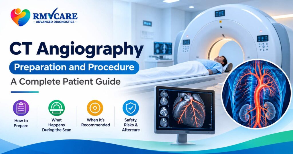
CT Angiography Preparation and Procedure: Everything You Need to Know

CT Angiography Preparation and Procedure: Everything You Need to Know When doctors need detailed images of blood vessels to investigate circulation-related conditions, they may recommend a specialised imaging examination called CT angiography (CTA). This advanced diagnostic technique combines computed tomography (CT) scanning with an iodine-based contrast material to produce detailed images of blood vessels and surrounding structures. Understanding CT angiography preparation and procedure can help patients feel more confident before their examination. Knowing what happens before, during and after the scan can also help you follow the correct instructions and avoid unnecessary delays. Whether your doctor has recommended a CT angiogram to examine blood vessels in the brain, heart, chest, abdomen or legs, this guide explains its purpose, preparation, procedure, benefits and important safety considerations. What Is CT Angiography? CT angiography, also known as a CT angiogram, is a specialised diagnostic imaging examination used to visualise blood vessels throughout the body. It uses X-rays, computer processing and an iodine-based contrast agent, usually injected into a vein, to create detailed images of arteries and selected vascular structures. Unlike conventional CT examinations that primarily focus on organs and tissues, CT angiography is specifically designed to evaluate blood vessels and identify certain abnormalities affecting circulation. Doctors may recommend this examination to investigate: The examination helps doctors understand the location and extent of certain vascular abnormalities and plan appropriate medical management. How Does CT Angiography Work? CT angiography works by combining CT imaging technology with intravenous contrast material. The contrast agent travels through the bloodstream and makes blood vessels more visible on the CT images. The scanner captures multiple images as the contrast moves through the targeted blood vessels. These images are then processed by specialised software to create detailed cross-sectional and, when appropriate, three-dimensional views. The radiologist can examine the images to assess the structure of blood vessels, identify possible narrowing or abnormalities and evaluate surrounding anatomical structures. What Are the Different Types of CT Angiography? CT angiography can examine blood vessels in different parts of the body. The specific examination depends on the doctor’s recommendation and the suspected medical condition. 1. CT Brain Angiography CT angiography of the brain examines blood vessels supplying the brain. It may be recommended to investigate certain aneurysms, vascular abnormalities, narrowed arteries and selected conditions affecting cerebral circulation. 2. CT Coronary Angiography CT coronary angiography, also called CCTA, examines the coronary arteries that supply blood to the heart. It may help doctors assess coronary artery plaque and narrowing in appropriately selected patients. 3. CT Pulmonary Angiography CT pulmonary angiography examines the blood vessels in the lungs. It is commonly used when doctors need to investigate a suspected pulmonary embolism, a blood clot affecting the pulmonary arteries. 4. CT Aortic Angiography This examination evaluates the aorta, the body’s largest artery. It may be recommended to investigate conditions such as aortic aneurysms, dissections and selected vascular abnormalities. 5. CT Peripheral Angiography CT peripheral angiography examines blood vessels in areas such as the legs and arms. It may help assess narrowing, blockages and other vascular abnormalities that affect blood circulation in the limbs. Why Do Doctors Recommend a CT Angiography? Doctors may recommend CT angiography when detailed information about blood vessels is required for diagnosis or treatment planning. Some common clinical applications include: The treating doctor determines whether CT angiography is appropriate based on symptoms, clinical history and other examination findings. CT Angiography Preparation: What Should You Do Before the Scan? Proper preparation is important for obtaining good-quality CT angiography images and ensuring patient safety. Preparation instructions may vary depending on the type of angiography, the contrast material used and your medical history. 1. Inform Your Doctor About Your Medical History Before the examination, inform your doctor and radiology team if you have: This information helps the medical team assess potential risks and plan the appropriate examination. 2. Follow Fasting Instructions Some CT angiography examinations require patients to avoid food or drinks for a specified period before the scan. The fasting duration can differ according to the examination and centre-specific protocol. Do not assume that every CT angiogram has the same fasting requirements. Confirm the exact instructions when scheduling your appointment. 3. Inform the Team About Your Medications Provide details of all medicines you currently take, including diabetes medication and supplements. Do not stop or change prescribed medicines without instructions from your treating doctor or radiology team. Certain examinations, particularly coronary CT angiography, may involve additional medication instructions depending on the patient’s heart rate and clinical condition. 4. Kidney Function Assessment Because CT angiography commonly involves iodine-based intravenous contrast, your doctor may request kidney function tests, such as serum creatinine or estimated glomerular filtration rate (eGFR). This is particularly important for patients with known kidney disease, acute kidney injury or other relevant risk factors. The radiology team will decide whether additional assessment or precautions are necessary. 5. Wear Comfortable Clothing Choose comfortable clothing without unnecessary metal objects. You may be asked to remove jewellery, watches, metal accessories or certain clothing items that could interfere with the images. 6. Inform the Team If You Are Pregnant or Breastfeeding If you are pregnant or think you may be pregnant, inform your doctor before undergoing CT imaging. If you are breastfeeding, discuss the use of contrast material with your healthcare professional so that you receive appropriate advice. CT Angiography Procedure: What Happens During the Scan? Understanding the CT angiography procedure can help reduce uncertainty before your appointment. Although the exact steps depend on the examination, the general process includes the following stages. Step 1: Patient Registration and Initial Assessment The healthcare team reviews your prescription, relevant medical history and preparation requirements. They may also check your previous reports and requested kidney function results, where applicable. Step 2: Positioning on the CT Scanner You will be asked to lie on the CT examination table. The radiology professional will position you according to the body area being examined to help obtain clear images. Step 3: Intravenous Cannula Placement A small intravenous cannula is usually

CT Angiography Preparation and Procedure: Everything You Need to Know Read More »
Doppler ultrasound

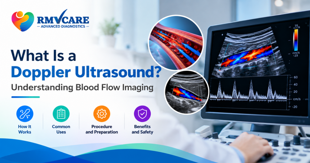
What Is a Doppler Ultrasound? Understanding Blood Flow Imaging

What Is a Doppler Ultrasound? Understanding Blood Flow Imaging Have you ever wondered how doctors check whether blood is flowing properly through your arteries and veins? A Doppler ultrasound is a specialised imaging test that helps evaluate blood circulation inside the body. It allows doctors to assess the direction and speed of blood flow and identify certain abnormalities affecting blood vessels. Unlike a conventional ultrasound that mainly produces images of internal structures, Doppler ultrasound provides additional information about moving blood cells. It is commonly used to investigate circulation-related concerns, assess blood vessels and monitor blood flow in selected organs. Understanding what a Doppler ultrasound is, how it works and when it may be recommended can help patients feel more prepared before undergoing the examination. What Is a Doppler Ultrasound? A Doppler ultrasound is a non-invasive diagnostic imaging technique that uses high-frequency sound waves to evaluate blood flow through blood vessels. The examination works on the Doppler effect, which measures changes in the frequency of sound waves reflected by moving red blood cells. A computer processes these changes to provide information about the direction and speed of blood circulation. Doctors may recommend Doppler ultrasound to examine arteries and veins in different parts of the body, including the neck, arms, legs and abdomen. It can help identify blood-flow abnormalities, narrowed blood vessels and certain circulation-related conditions. How Does a Doppler Ultrasound Work? A Doppler ultrasound examination uses a handheld device called a transducer. The transducer sends sound waves into the body and receives the echoes reflected by moving blood cells. The ultrasound system analyses these echoes and converts the information into visual images, graphs or colour-coded displays that help assess blood circulation. Here is how the examination generally works: The procedure is generally painless and does not use ionising radiation. What Are the Different Types of Doppler Ultrasound? Different Doppler techniques provide different information about blood circulation. The appropriate method depends on the area being examined and the medical question. 1. Colour Doppler Ultrasound Colour Doppler uses colour-coded information to display the direction and relative characteristics of blood flow within blood vessels. It helps doctors assess circulation patterns and identify certain abnormalities in blood flow. 2. Power Doppler Ultrasound Power Doppler is particularly sensitive to detecting relatively slow or small-volume blood flow in selected situations. It can help evaluate blood flow in smaller vessels and certain organs. However, it does not provide the direction of blood flow in the same way as colour Doppler. 3. Spectral Doppler Ultrasound Spectral Doppler displays blood-flow measurements as graphs over time. It helps assess the speed and pattern of blood circulation through a vessel. Doctors may use this information to investigate abnormal flow patterns or narrowing in blood vessels. 4. Duplex Doppler Ultrasound Duplex ultrasound combines conventional ultrasound images with Doppler blood-flow measurements. It allows doctors to examine both the structure of blood vessels and the movement of blood through them. Why Do Doctors Recommend a Doppler Ultrasound? A Doppler ultrasound may be recommended when a doctor needs more information about blood circulation in a particular part of the body. Some common clinical applications include: 1. Detecting Blood Clots A venous Doppler ultrasound is commonly used to investigate suspected deep vein thrombosis (DVT), a condition involving blood clots in deep veins, particularly in the legs. The examination helps assess blood flow and identify findings that may indicate a clot. 2. Evaluating Narrowed or Blocked Arteries Arterial Doppler examinations help assess blood circulation through arteries. They may be recommended when a doctor suspects narrowing or reduced blood flow. 3. Investigating Leg Swelling and Circulation Problems Unexplained leg swelling, discomfort or certain circulation-related symptoms may lead a doctor to recommend a venous or arterial Doppler examination. The type of scan depends on the suspected cause. 4. Assessing Carotid Arteries A carotid Doppler ultrasound examines the major arteries in the neck that supply blood to the brain. It may help assess plaque buildup, narrowing and blood-flow abnormalities in these vessels. 5. Evaluating Blood Flow to Certain Organs Doppler ultrasound may also be used to assess blood circulation in selected organs, including the kidneys and liver, when clinically indicated. 6. Monitoring Blood Flow During Pregnancy Certain obstetric Doppler examinations help evaluate blood flow between the mother, placenta and developing baby. These scans are recommended by doctors when additional information about fetal or placental circulation is needed. Which Body Parts Can Be Examined Using Doppler Ultrasound? Depending on the doctor’s recommendation and the available examination protocol, Doppler ultrasound can evaluate blood flow in different areas. Examination What it evaluates Venous Doppler Blood flow through selected veins, commonly in the legs Arterial Doppler Blood circulation through arteries Carotid Doppler Blood flow through arteries in the neck Renal Doppler Blood circulation in the kidneys Hepatic Doppler Blood flow through selected liver blood vessels Obstetric Doppler Selected blood-flow patterns involving the pregnancy Not every Doppler examination is suitable for every patient. Your doctor will recommend the appropriate scan based on your symptoms and clinical history. How to Prepare for a Doppler Ultrasound? Preparation depends on the type of Doppler ultrasound and the body part being examined. Some examinations require little or no preparation, while others may involve specific instructions. Before your appointment, consider the following: Always confirm the preparation requirements for your particular examination before visiting the diagnostic centre. What Happens During a Doppler Ultrasound Examination? A Doppler ultrasound is generally performed while the patient is lying down or sitting in a comfortable position, depending on the area being examined. The sonographer applies ultrasound gel to the skin and gently moves the transducer over the examination area. The machine captures images and evaluates blood-flow patterns. You may hear soft whooshing sounds from the equipment as it processes the Doppler signals. The examination is usually painless, although mild pressure from the transducer may occasionally cause temporary discomfort, particularly if the area is already tender. Once the examination is complete, the gel is removed and the images are reviewed for reporting. The duration depends on

What Is a Doppler Ultrasound? Understanding Blood Flow Imaging Read More »

liver infection symptoms

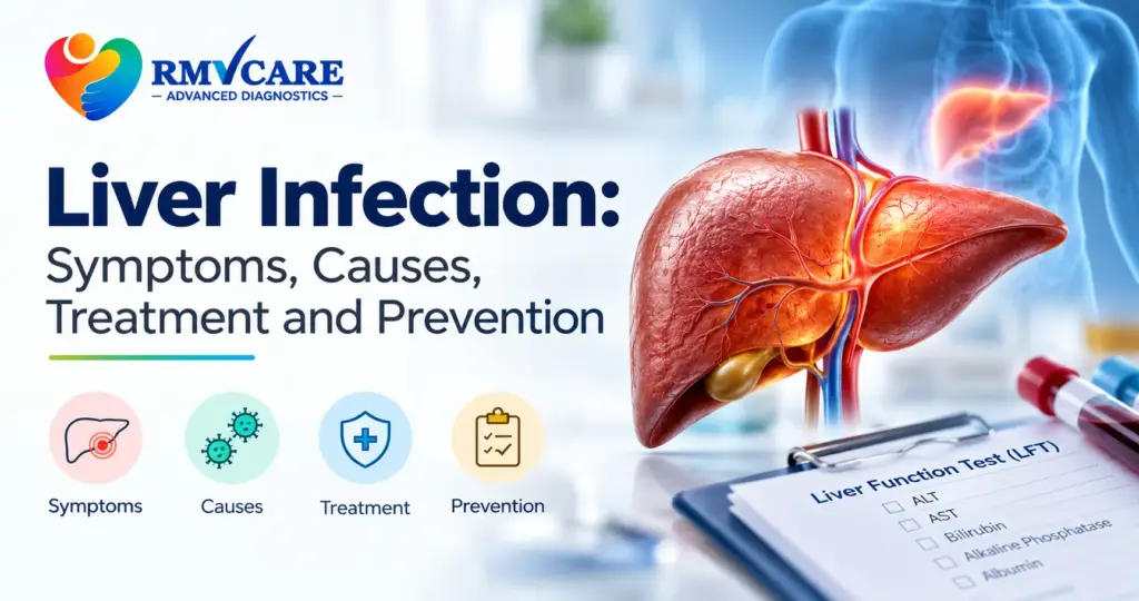
Liver Infection: Symptoms, Causes, Treatment and Prevention

Liver Infection: Symptoms, Causes, Treatment and Prevention The liver is one of the most important organs in the human body. It helps process nutrients, produces bile for digestion, stores energy and removes harmful substances from the blood. When the liver becomes inflamed or infected, its normal functions may be affected. Understanding liver infection symptoms, possible causes and available treatment options can help people seek medical attention at the right time. A liver infection is commonly associated with viral hepatitis, although other infections and medical conditions can also affect the liver. Some people experience symptoms such as fatigue, nausea, abdominal discomfort and yellowing of the skin or eyes, while others may have no noticeable symptoms during the early stages. In this guide, we explain the common symptoms of liver infection, its causes, different types of hepatitis, diagnostic tests, treatment options and practical prevention measures. What Is a Liver Infection? A liver infection occurs when an infectious organism, most commonly a virus, affects the liver and causes inflammation or damage. Viral hepatitis is one of the most recognised forms of liver infection. The five main hepatitis viruses are hepatitis A, B, C, D and E. Each type spreads differently and may have different effects on the liver. Some infections are usually short-term, while others can become chronic and increase the risk of long-term liver damage. It is important to understand that not every liver condition is an infection. Fatty liver disease, alcohol-related liver disease, autoimmune hepatitis and certain medication-related injuries can also cause liver inflammation or damage without a contagious infection. Common Symptoms of Liver Infection The symptoms of a liver infection can vary depending on the type of infection, its severity and the person’s overall health. Some infections remain asymptomatic for months or years. Common liver infection symptoms include: 1. Yellowing of the Skin and Eyes Yellowing of the skin or the whites of the eyes is known as jaundice. It occurs when bilirubin builds up in the body. Jaundice can be associated with hepatitis and other liver or bile duct conditions and requires prompt medical assessment. 2. Dark-Coloured Urine Urine that appears darker than usual, such as tea-coloured urine, may occur when bilirubin levels increase. Dark urine can have other causes too, so it should be evaluated alongside other symptoms. 3. Unusual Tiredness and Weakness Persistent fatigue, weakness or a lack of energy can occur during a liver infection. If tiredness continues without an obvious explanation, consult a healthcare professional rather than assuming it is caused by a liver problem. 4. Loss of Appetite Some people experience reduced appetite, nausea or a general feeling of being unwell. These symptoms may make it difficult to eat normally and can sometimes lead to weight loss. 5. Nausea and Vomiting Nausea and vomiting can occur with acute hepatitis and other illnesses affecting the digestive system. Persistent vomiting may cause dehydration and should not be ignored. 6. Abdominal Pain or Discomfort Discomfort in the upper-right part of the abdomen, beneath the ribs, can sometimes occur when the liver is inflamed. However, abdominal pain has many possible causes and cannot confirm a liver infection on its own. 7. Pale or Clay-Coloured Stools Stools that become unusually pale may indicate a problem with bile flow. This symptom, particularly when accompanied by jaundice or dark urine, needs prompt medical evaluation. 8. Fever and Joint Pain Some viral infections can cause fever, body aches, joint pain or flu-like symptoms before other signs of hepatitis appear. When should you seek medical help? Seek prompt medical assessment if you notice yellowing of the eyes or skin, persistent dark urine with pale stools, or unexplained abdominal discomfort. Severe confusion, unusual drowsiness, severe abdominal pain, repeated vomiting or bleeding require emergency medical attention. What Causes a Liver Infection? The causes of a liver infection depend on the type of organism involved and how it enters the body. 1. Viral Hepatitis Hepatitis viruses are a major cause of liver infections. Each type has different transmission routes and health risks. 2. Contaminated Food and Water Unsafe drinking water, contaminated food and poor sanitation can increase the risk of hepatitis A and E. Washing hands properly, consuming safe drinking water and following food hygiene practices are important preventive measures. 3. Unsafe Medical or Personal-Care Equipment Equipment contaminated with infected blood can transmit certain hepatitis viruses. Examples include improperly sterilised medical instruments, reused needles and unsafe tattooing or piercing equipment. Choose qualified healthcare providers and facilities that follow appropriate infection-control and sterilisation procedures. 4. Contact With Infected Blood Sharing needles, razors or other items contaminated with infected blood may expose a person to hepatitis viruses. Hepatitis B can also spread through certain sexual contacts and from an infected mother to her baby during childbirth. Hepatitis viruses do not all spread in the same way. Casual contact, such as sitting beside someone or sharing an ordinary conversation, is not a typical route of transmission for hepatitis B or C. 5. Other Conditions That Affect the Liver Not every condition described as liver inflammation is an infection. Excessive alcohol consumption, fatty liver disease, autoimmune disorders and some medicines can damage the liver. A healthcare professional can help determine the actual cause. Types of Liver Infection Understanding the different types of viral hepatitis is important because diagnosis, treatment and prevention depend on the specific virus. Hepatitis A Hepatitis A usually causes an acute infection and does not become chronic. Many people recover with supportive care, including adequate fluids, rest and appropriate nutrition. Vaccination can help prevent infection. Hepatitis B Hepatitis B can cause either acute or chronic infection. Chronic hepatitis B may damage the liver over time and increase the risk of cirrhosis and liver cancer. Antiviral medicines are available for people who meet treatment criteria, and vaccination provides effective protection against infection. Hepatitis C Hepatitis C is primarily transmitted through infected blood. It may remain undetected for years because symptoms are often absent. Modern antiviral medicines can cure most hepatitis C infections, making timely testing and medical

Liver Infection: Symptoms, Causes, Treatment and Prevention Read More »

6 Body Signals That May Appear Before a Heart Attack

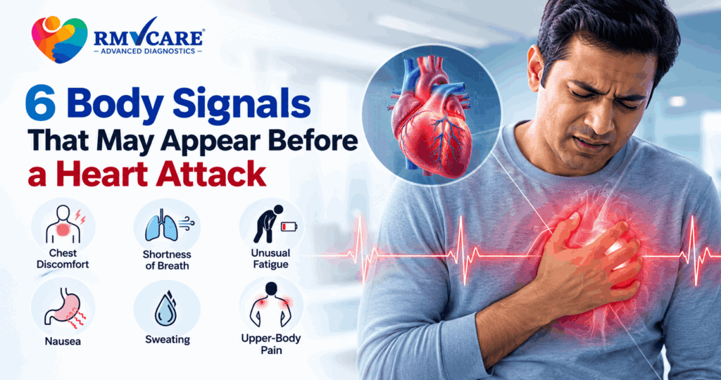
6 Body Signals That May Appear Before a Heart Attack

6 Body Signals That May Appear Before a Heart Attack A heart attack can happen suddenly, but the body may sometimes show warning symptoms that should not be ignored. Recognising these heart attack warning signs can help a person seek medical attention quickly. A heart attack occurs when blood flow to part of the heart muscle becomes blocked or significantly reduced. Without timely treatment, the heart muscle can become damaged. www.heart.org Not everyone experiences the same symptoms. Some people may have significant chest discomfort, while others may mainly experience breathlessness, nausea, unusual tiredness or pain in another part of the upper body. Here are 6 important body signals that may indicate a heart problem or possible heart attack. 1. Chest Pressure, Tightness or Discomfort One of the most recognised signs of a heart attack is discomfort in the centre of the chest. It may feel like: Chest discomfort should not automatically be assumed to be acidity or indigestion, particularly when it is unusual, persistent or accompanied by other symptoms. The American Heart Association notes that heart attack-related chest discomfort may last more than a few minutes or may disappear and return. www.heart.org Do not ignore new or unexplained chest discomfort. Seek urgent medical evaluation. 2. Pain or Discomfort in the Arm, Shoulder, Jaw, Neck or Back Heart-related discomfort does not always remain in the chest. Pain or discomfort may spread to other parts of the upper body, including: This is one reason heart attack symptoms can sometimes be mistaken for muscle pain, dental problems or other common conditions. According to the American Heart Association, discomfort involving the arms, back, neck, jaw or stomach can occur during a heart attack. www.heart.org If unexplained upper-body discomfort occurs together with chest pressure, breathlessness, sweating or nausea, it should be treated as a potential medical emergency. 3. Shortness of Breath Sudden or unusual shortness of breath can be another warning sign. You may feel: Importantly, shortness of breath can occur with or without chest discomfort during a heart attack. www.heart.org Breathlessness has many possible causes, so it does not automatically mean that someone is having a heart attack. However, sudden or unexplained breathing difficulty—especially when accompanied by other symptoms—requires prompt medical attention. 4. Unusual Tiredness, Weakness or Lightheadedness Feeling extremely tired without an obvious reason can sometimes accompany heart-related problems. Some people may experience: These symptoms can easily be attributed to stress, lack of sleep or a busy routine. However, when unusual fatigue or lightheadedness occurs together with chest discomfort, breathlessness, nausea or sweating, it should not be ignored. The American Heart Association lists unusual tiredness, lightheadedness and feeling faint among possible heart attack symptoms. www.heart.org 5. Nausea, Vomiting or Indigestion-Like Discomfort Heart attack symptoms can sometimes resemble digestive problems. A person may experience: This can make it difficult to recognise a heart attack, particularly when there is little or no obvious chest pain. Nausea and vomiting are recognised possible symptoms of a heart attack, and women may be more likely to experience symptoms such as nausea, unusual fatigue, shortness of breath or back and shoulder discomfort. www.heart.org If digestive-like symptoms are sudden, unusual or accompanied by other warning signs, medical assessment should not be delayed. 6. Cold Sweating or Sudden Sweating Breaking out into a cold sweat without an obvious reason can be another warning sign. For example, if someone suddenly becomes sweaty while experiencing: it is important to seek urgent medical attention. Cold sweating is recognised as one of the possible warning signs associated with heart attack. www.heart.org Can Heart Attack Symptoms Be Different in Women? Yes. Although chest pain or discomfort remains a common heart attack symptom in women, women can also experience symptoms that may appear less typical. These can include: Because some of these symptoms can resemble everyday health problems, they may sometimes be overlooked. www.heart.org This is why unusual or sudden symptoms should be taken seriously, particularly when several symptoms occur together. Are These Always Signs That a Heart Attack Is About to Happen? No. These symptoms can have many different causes and do not necessarily mean that a person will have a heart attack. For example, chest discomfort can occur because of digestive problems, muscle strain or other medical conditions. Similarly, fatigue and breathlessness can have numerous causes. However, because heart attacks can be life-threatening, new, severe or unexplained symptoms should not be self-diagnosed. A heart attack can also occur without obvious warning symptoms. Some people experience a “silent” heart attack with mild or unusual symptoms that may not immediately be recognised. www.heart.org What Should You Do If You Notice These Symptoms? If you or someone around you develops symptoms that could indicate a heart attack, do not wait to see whether they disappear. Seek emergency medical care immediately, especially when symptoms include chest pressure or discomfort, difficulty breathing, pain spreading to the arm, jaw, neck or back, sweating, nausea or faintness. www.heart.org Do not attempt to diagnose the problem based only on an online article. Every minute can matter during a heart attack. How Can Heart Health Be Monitored? 6 Body Signals That May Appear Before a Heart Attack Regular health assessments can help identify cardiovascular risk factors and provide useful information about heart health. Depending on your symptoms, medical history and risk factors, a healthcare professional may recommend investigations such as: ECG An ECG (electrocardiogram) records the electrical activity of the heart and can help healthcare professionals assess certain heart-related problems. 2D Echo A 2D echocardiogram uses ultrasound to create images of the heart and helps assess its structure and function. Stress Test A cardiac stress test evaluates how the heart responds to increased physical activity and may be recommended in selected patients based on symptoms and clinical assessment. The appropriate test depends on the individual’s symptoms and medical history. A diagnostic test should always be recommended and interpreted by a qualified healthcare professional. Who Should Pay More Attention to Heart Health? Heart health deserves particular attention if you have risk factors such as:

6 Body Signals That May Appear Before a Heart Attack Read More »

DEXA scan for osteoporosis

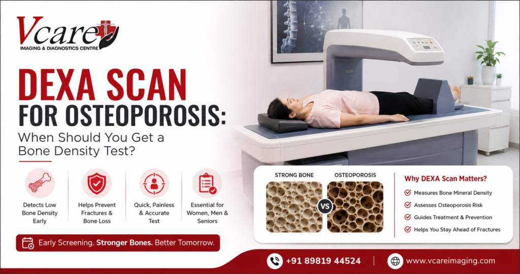
DEXA Scan for Osteoporosis: When Should You Get a Bone Density Test?

DEXA Scan for Osteoporosis: When Should You Get a Bone Density Test? Osteoporosis can develop silently, weakening bones without causing noticeable symptoms until a fracture occurs. A DEXA scan for osteoporosis is one of the most commonly used tests to measure bone mineral density and help identify low bone density and osteoporosis before a serious fracture happens. But when should you get a DEXA scan? Is a bone density test only recommended for older adults, or can younger people also need one? Understanding when bone density testing is appropriate can help you take timely steps to protect your bone health. In this guide, VCare Imaging and Diagnostic Centre explains who may need a DEXA scan for bone density, how the test works, what the results mean, and when your doctor may recommend osteoporosis screening. What Is a DEXA Scan? A DEXA scan, also known as a dual-energy X-ray absorptiometry scan, is a specialized imaging test used to measure bone mineral density (BMD). A bone density test commonly examines areas such as the hip and lower spine because these locations are particularly important when assessing fracture risk. Unlike a standard X-ray, a DEXA scan is designed specifically to measure bone density. The results can help doctors identify whether your bone density is within the expected range or whether you may have osteopenia or osteoporosis. What Is Osteoporosis? Osteoporosis is a condition in which bones become weaker and more fragile, increasing the risk of fractures. Bone tissue is constantly being broken down and rebuilt. As people age, bone loss can gradually become greater than new bone formation. This can reduce bone density and make bones more susceptible to fractures. Osteoporosis often develops without obvious symptoms. For this reason, osteoporosis screening and appropriate bone density testing can be important for people who have risk factors. How Does a DEXA Scan Help Detect Osteoporosis? A DEXA scan for osteoporosis measures bone mineral density and provides a numerical score that doctors can use when assessing bone health. The test commonly provides a T-score, which compares your bone density with that of a healthy young adult reference population. Generally: Your doctor will interpret your DEXA scan results along with your age, medical history, risk factors and other clinical information. When Should You Get a Bone Density Test? The appropriate time for a bone density test for osteoporosis depends on your age, sex, health history and risk factors. A doctor may recommend a DEXA scan if you have an increased risk of bone loss or fractures. 1. Women After Menopause Bone loss can accelerate after menopause because of hormonal changes, particularly reduced estrogen levels. For this reason, postmenopausal women may be advised to discuss osteoporosis screening and DEXA testing with their healthcare provider. 2. Older Adults The risk of osteoporosis generally increases with age. Older adults may be recommended a DEXA scan for bone density as part of osteoporosis screening, particularly when other risk factors are present. 3. People With a Previous Fragility Fracture A fracture resulting from a relatively minor fall or injury can sometimes indicate reduced bone strength. If you have experienced a fragility fracture, your doctor may recommend a bone density test to assess your risk of osteoporosis and future fractures. 4. People With a Family History of Osteoporosis Genetics can influence bone health. A family history of osteoporosis or hip fracture may increase your risk. If osteoporosis runs in your family, discuss your individual risk with your healthcare provider to determine whether a DEXA scan is appropriate. 5. People With Certain Medical Conditions Some health conditions can contribute to reduced bone density. Depending on your medical history, your doctor may recommend a bone density test if you have a condition that may affect bone metabolism or increase fracture risk. 6. People Taking Certain Long-Term Medications Some medications can affect bone health when used for prolonged periods. For example, long-term corticosteroid treatment can increase the risk of bone loss. Your doctor may recommend a DEXA scan depending on the medication, dose, duration and your other risk factors. 7. People With Low Body Weight or Other Risk Factors Low body weight and certain lifestyle or nutritional factors may be associated with an increased risk of low bone density. If you have multiple osteoporosis risk factors, your healthcare provider may recommend bone density testing even if you have not experienced a fracture. What Happens During a DEXA Scan? A DEXA scan is generally a quick and non-invasive examination. During the procedure: The procedure is generally painless, and there is usually no recovery period required after the examination. How Long Does a DEXA Scan Take? A DEXA scan is typically a relatively quick examination. The exact duration can vary depending on the areas being scanned and the imaging center’s protocol. Your diagnostic center can provide specific information about the expected examination time and preparation requirements when you schedule your appointment. How to Prepare for a DEXA Scan Preparation requirements can vary depending on the imaging center and your individual circumstances. Before your appointment, you may be advised to: Always follow the instructions provided by your healthcare provider or imaging center. What Do DEXA Scan Results Mean? The main measurement used to assess osteoporosis in many adults is the T-score. Understanding the DEXA Scan T-Score The T-score indicates how your bone mineral density compares with the bone density of a healthy young adult reference population. A lower T-score generally indicates lower bone density. Your doctor may use the T-score along with other clinical factors to determine whether you have normal bone density, osteopenia or osteoporosis. What Is a Z-Score? A Z-score compares your bone density with that of people of similar age, sex and other relevant characteristics. Z-scores can be particularly useful in certain younger individuals and specific clinical situations. A healthcare professional should interpret your DEXA scan results rather than relying on the score alone. Osteopenia vs Osteoporosis: What Is the Difference? Osteopenia and osteoporosis both involve reduced bone density, but they are not the same.

DEXA Scan for Osteoporosis: When Should You Get a Bone Density Test? Read More »

DEXA scan

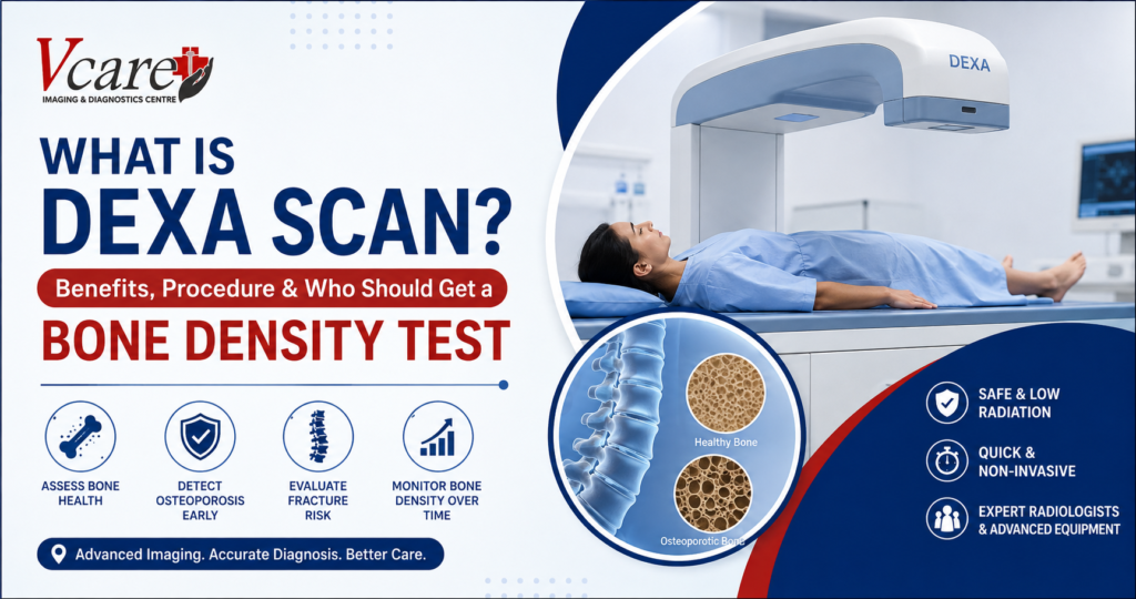
What Is DEXA Scan? Benefits, Procedure & Who Should Get a Bone Density Test

What Is a DEXA Scan? Benefits, Procedure & Who Should Get a Bone Density Test A DEXA scan is a specialized imaging test used to measure bone mineral density and assess bone health. DEXA stands for Dual-Energy X-ray Absorptiometry and is also commonly called a DXA scan or bone density test. The test uses a very small amount of X-ray radiation to measure bone mineral density, usually at the hip and lower spine. It is widely used to help diagnose osteoporosis, assess fracture risk, and monitor changes in bone density over time. A bone density test can be particularly useful when there are concerns about weakened bones or an increased risk of osteoporosis. Your doctor may recommend a DEXA scan based on your age, medical history, previous fractures, medications, or other risk factors. If you have been advised to undergo a DEXA scan in Kandivali, understanding the procedure and what the results mean can help you feel more prepared. Why Is a Bone Density Test Important? Bones naturally change throughout life. Bone loss can become more significant with age and may increase the risk of fractures. Osteoporosis is a condition in which bones become weaker and more fragile, increasing the likelihood of fractures. Because bone loss can develop without obvious symptoms, a bone density test can help identify reduced bone mineral density before a serious fracture occurs. A DEXA scan can help healthcare professionals: A DEXA scan does not predict with certainty whether an individual will experience a fracture. Instead, the results are considered along with other clinical risk factors to help doctors assess bone health and decide on appropriate management. What Does a DEXA Scan Measure? A DEXA bone density scan measures bone mineral density, commonly abbreviated as BMD. During the test, low-dose X-rays are used to measure the amount of X-ray energy absorbed by bone and soft tissue. The information is processed to calculate bone density measurements. The most commonly examined areas are: The hip and spine are commonly used because these areas are important for assessing bone health and fracture risk. 7 Benefits of a DEXA Scan 1. Helps Measure Bone Mineral Density The primary purpose of a DEXA scan is to measure bone mineral density. This provides doctors with objective information about the strength and mineral content of bones and can help identify low bone density. 2. Helps Detect Osteoporosis A DEXA scan is one of the standard imaging methods used to diagnose osteoporosis. It can identify reduced bone density and help healthcare professionals assess whether further evaluation or treatment may be appropriate. Early identification can be valuable because osteoporosis may progress without noticeable symptoms until a fracture occurs. 3. Helps Assess Fracture Risk Low bone density is associated with an increased risk of osteoporotic fractures. DEXA results can be considered alongside factors such as age, previous fractures, family history, body weight, lifestyle factors, and certain medical conditions when assessing fracture risk. 4. Supports Monitoring of Bone Health A bone density test may also be used to monitor changes in bone density over time. For people receiving treatment for osteoporosis or other conditions associated with bone loss, follow-up DEXA scans may help doctors evaluate changes and treatment response. 5. Quick and Non-Invasive A DEXA scan is generally a quick, non-invasive imaging examination. No anesthesia is normally required, and the patient typically lies on a padded examination table while the scanner obtains measurements. 6. Uses a Very Low Radiation Dose DEXA uses a small amount of ionizing radiation. The radiation exposure is considerably lower than that of many conventional X-ray examinations. Even so, always inform your doctor and the imaging technologist if you are pregnant or may be pregnant. 7. Helps Doctors Make Informed Decisions A DEXA scan provides measurable information about bone mineral density. When interpreted alongside your medical history and other risk factors, the results can help your healthcare provider make informed decisions about further evaluation, monitoring, or treatment. Who Should Get a DEXA Scan? Not everyone needs a DEXA scan routinely. The decision depends on individual risk factors and your doctor’s recommendation. A bone density test may be considered for people who have an increased risk of osteoporosis or bone loss. Your doctor may recommend a DEXA scan if you have: Postmenopausal women and older adults may have an increased risk of osteoporosis, but bone loss can also affect men and younger individuals in certain circumstances. The appropriate timing of a DEXA bone density scan should therefore be based on individual risk factors and medical advice. DEXA Scan for Women Bone density can decline after menopause, making bone health an important consideration for women as they age. A doctor may recommend a DEXA scan for women based on age, menopause status, fracture history, family history, medications, and other risk factors. Women who have additional risk factors for osteoporosis may need assessment earlier than someone without those risk factors. If you are concerned about bone health after menopause, discuss your individual risk factors with your doctor to determine whether a bone density test is appropriate. DEXA Scan for Men Osteoporosis is not limited to women. Men can also develop low bone density, particularly when certain medical conditions, medications, lifestyle factors, or age-related changes increase their risk. A doctor may recommend a DEXA scan for men when there are specific risk factors for osteoporosis or bone loss. What Happens During a DEXA Scan Procedure? Understanding the DEXA scan procedure can make the experience easier for first-time patients. Step 1: Patient Preparation Before the examination, the imaging team may ask about your medical history, medications, previous imaging examinations, and any relevant health conditions. You should tell your doctor or technologist if you may be pregnant. Step 2: Positioning You will usually lie on a padded examination table. For a central DEXA examination, the scanner generally evaluates the hip and lower spine. Your position may be adjusted so that the required areas can be accurately assessed. Step 3: Scanning The DEXA machine passes a scanning device over

What Is DEXA Scan? Benefits, Procedure & Who Should Get a Bone Density Test Read More »

Fatty Liver Diagnosis

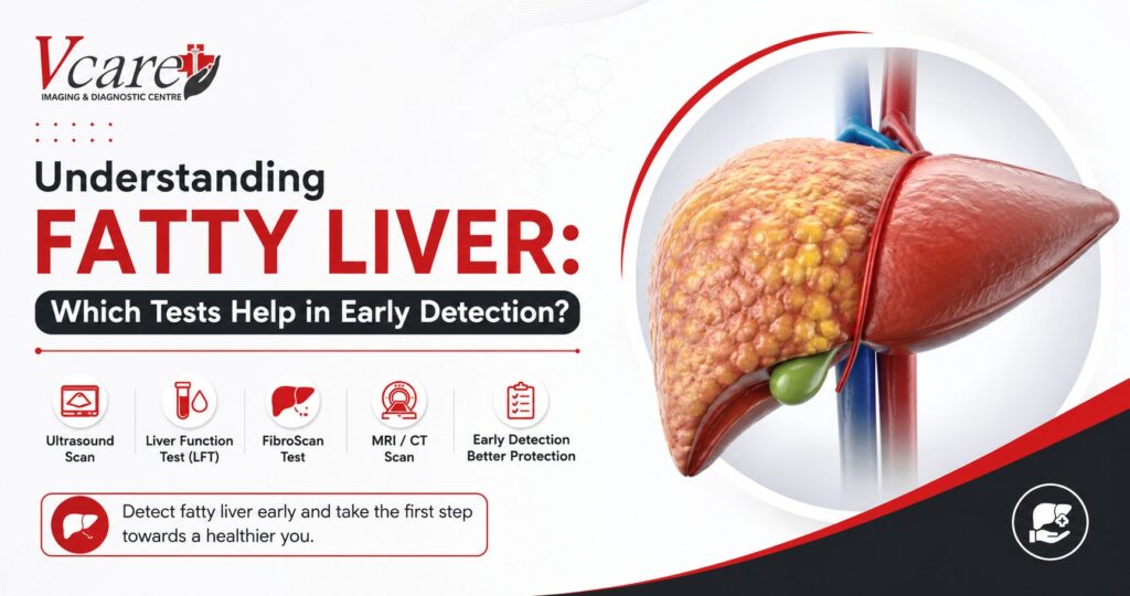
Understanding Fatty Liver: Which Tests Help in Early Detection?

Understanding Fatty Liver: Which Tests Help in Early Detection? Fatty liver disease has become one of the most common lifestyle-related health conditions worldwide. It often develops silently, with little or no noticeable symptoms during its early stages. Because of this, many people remain unaware of the condition until it progresses into more serious liver damage. An accurate fatty liver diagnosis plays a vital role in detecting the condition early, allowing timely treatment and lifestyle modifications that can help prevent complications such as liver fibrosis, cirrhosis, and even liver failure. This guide explains everything you need to know about fatty liver diagnosis, including symptoms, causes, diagnostic tests, and when you should consider getting screened. What Is Fatty Liver Disease? Fatty liver disease occurs when excess fat accumulates inside the liver cells. While a small amount of fat is normal, excessive fat can affect liver function and increase the risk of inflammation and long-term liver damage. There are two primary types of fatty liver disease: Among these, NAFLD is becoming increasingly common due to unhealthy eating habits, obesity, diabetes, and sedentary lifestyles. Common Symptoms of Fatty Liver In many cases, fatty liver disease does not cause noticeable symptoms. However, as the condition progresses, patients may experience: Since these symptoms are often mild or absent, fatty liver diagnosis through regular health screenings becomes essential. Who Is at Risk? Certain individuals have a higher risk of developing fatty liver disease. You may be at higher risk if you have: Routine fatty liver diagnosis is recommended for individuals with these risk factors. Which Tests Help in Fatty Liver Diagnosis? Several diagnostic tests help doctors evaluate liver health and determine the severity of fatty liver disease. 1. Liver Function Test (LFT) A Liver Function Test measures enzymes and proteins in your blood that indicate how well your liver is functioning. Although an LFT cannot confirm fatty liver by itself, abnormal liver enzyme levels often prompt further investigations. 2. Ultrasound Scan Ultrasound is one of the most commonly recommended imaging tests for fatty liver diagnosis. It is: An abdominal ultrasound can identify fat deposits inside the liver before symptoms become severe. 3. FibroScan (Liver Elastography) FibroScan is an advanced, non-invasive test that measures liver stiffness and fat content. Doctors recommend FibroScan to: 4. CT Scan A CT scan provides detailed cross-sectional images of the liver and surrounding organs. While not always the first choice for fatty liver diagnosis, it can help detect liver abnormalities when additional imaging is required. 5. MRI Scan MRI offers highly detailed liver imaging and is particularly useful for accurately measuring liver fat content. It is often recommended in complex cases or when precise evaluation is needed. 6. Blood Tests Doctors may also recommend additional blood investigations, including: These tests help identify underlying conditions contributing to fatty liver disease. Why Early Fatty Liver Diagnosis Matters Early detection significantly improves treatment outcomes. Benefits include: The earlier the fatty liver diagnosis, the easier it is to manage the condition. Can Fatty Liver Be Reversed? Yes. In many cases, early-stage fatty liver disease is reversible. Doctors usually recommend: Regular follow-up testing helps monitor liver health and treatment progress. When Should You Get Screened? You should consider a fatty liver diagnosis if you: Routine health screenings can detect fatty liver before serious complications develop. Why Choose VCare Imaging & Diagnostic Centre? At VCare Imaging & Diagnostic Centre, we provide advanced diagnostic services to support accurate fatty liver diagnosis. Our services include: Our goal is to help patients detect liver conditions early for timely treatment and improved health outcomes. Conclusion Fatty liver disease often develops silently, making regular health screenings essential for early detection. An accurate fatty liver diagnosis using imaging tests and blood investigations allows doctors to identify the condition before permanent liver damage occurs. If you have risk factors such as obesity, diabetes, or high cholesterol, don’t wait for symptoms to appear. Early diagnosis and timely lifestyle changes can protect your liver and improve your overall well-being. Schedule your liver screening today and take the first step toward better liver health. Frequently Asked Questions (FAQs)

Understanding Fatty Liver: Which Tests Help in Early Detection? Read More »

Monsoon Health Screening for preventive healthcare

Monsoon Health Screening: Why Preventive Diagnostic Tests Matter More During the Rainy Season

Monsoon Health Screening: Why Preventive Diagnostic Tests Matter More During the Rainy Season The arrival of the monsoon season brings welcome relief from the summer heat, lush greenery, and refreshing rainfall. However, along with the pleasant weather comes an increased risk of infections, waterborne diseases, and seasonal health complications. From viral fevers and respiratory infections to dengue and malaria, monsoon-related illnesses can affect people of all ages. This is where Monsoon Health Screening becomes essential. A proactive approach to health through timely diagnostic tests can help identify potential health concerns before they become serious. Instead of waiting for symptoms to worsen, preventive health screenings allow individuals to stay informed about their health and take necessary precautions during the rainy season. In this blog, we’ll explore why a Monsoon Health Screening is important, the key diagnostic tests to consider, and how preventive healthcare can help you enjoy a healthier monsoon. Why Is Health Risk Higher During the Monsoon? The rainy season creates favorable conditions for the growth of bacteria, viruses, and mosquitoes. Stagnant water becomes a breeding ground for disease-carrying insects, while increased humidity can contribute to respiratory and fungal infections. Common health concerns during monsoon include: Many of these conditions initially present with mild symptoms that can easily be overlooked. Early diagnosis through a Monsoon Health Screening can help detect underlying infections and prevent complications. What Is a Monsoon Health Screening? A Monsoon Health Screening is a preventive health assessment designed to evaluate your overall health and identify any early signs of infection or illness commonly associated with the rainy season. These screenings typically include blood tests, urine tests, and other diagnostic evaluations that provide valuable insights into your health status. The goal is to detect abnormalities before they lead to severe health issues. Preventive healthcare is not only about treating illnesses but also about preventing them. By undergoing a Preventive Health Checkup during monsoon, individuals can make informed decisions regarding their health and lifestyle. Benefits of Monsoon Health Screening 1. Early Detection of Seasonal Diseases Many monsoon-related diseases begin with common symptoms such as fever, fatigue, headache, and body pain. Diagnostic tests help determine the exact cause and ensure timely treatment. Early detection can significantly reduce the risk of complications associated with diseases like dengue, malaria, and typhoid. 2. Monitoring Overall Health A Health Checkup in Monsoon does more than detect infections. It provides a comprehensive overview of important health parameters, including blood sugar levels, liver function, kidney function, and immune response. Regular monitoring helps individuals maintain optimal health throughout the season. 3. Preventing Serious Health Complications Ignoring early symptoms can lead to delayed diagnosis and prolonged recovery. Preventive screenings identify potential concerns before they become severe, allowing for prompt medical intervention. 4. Peace of Mind Knowing that your health parameters are within a healthy range offers reassurance and confidence during a season known for increased health risks.\ Essential Diagnostic Tests During Monsoon A comprehensive Monsoon Health Screening may include several important tests depending on individual health needs and risk factors. Complete Blood Count (CBC) A CBC test helps evaluate overall health and can detect infections, anemia, and abnormalities in blood cells. It is often one of the first tests recommended when experiencing fever or fatigue. Dengue Profile Test Dengue cases tend to rise during monsoon due to mosquito breeding. A dengue profile test helps identify the infection early and monitor platelet levels. Malaria Test Malaria remains a common concern during the rainy season. Early detection through diagnostic testing enables timely treatment and prevents complications. Typhoid Test Contaminated food and water can increase the risk of typhoid during monsoon. Testing helps confirm infection and guide appropriate medical care. Liver Function Test (LFT) Certain infections can affect liver health. Liver function tests assess how well the liver is working and help identify any abnormalities. Kidney Function Test (KFT) Kidney function tests evaluate the health of the kidneys and ensure they are functioning properly. Urine Analysis Urine testing can detect infections, dehydration, and other health conditions that may require medical attention. Chest X-Ray (If Recommended) For individuals experiencing persistent cough, breathing difficulties, or respiratory symptoms, a chest X-ray may help identify underlying lung conditions. Who Should Consider a Monsoon Health Screening? While everyone can benefit from preventive healthcare, certain groups should prioritize Diagnostic Tests During Monsoon: Children Children are more vulnerable to infections due to their developing immune systems. Senior Citizens Older adults often have weaker immunity and may be at higher risk of seasonal illnesses. Individuals with Chronic Conditions People with diabetes, hypertension, asthma, or heart disease should consider regular health monitoring during monsoon. Working Professionals Busy schedules often lead to delayed medical attention. Preventive screenings help identify concerns before they affect productivity and well-being. Tips to Stay Healthy During Monsoon Along with regular Monsoon Health Screening, adopting healthy habits can help reduce the risk of illness. Maintain Proper Hygiene Wash your hands frequently and avoid consuming unhygienic food. Drink Safe Water Always drink filtered or boiled water to reduce the risk of waterborne diseases. Eat Fresh and Nutritious Food A balanced diet rich in fruits, vegetables, and essential nutrients supports a strong immune system. Prevent Mosquito Breeding Remove stagnant water around your home and use mosquito repellents when necessary. Stay Physically Active Regular exercise helps strengthen immunity and improve overall health. Seek Medical Advice Early Do not ignore symptoms such as persistent fever, fatigue, or unusual discomfort. Early consultation and diagnostic testing can make a significant difference. Why Choose a Trusted Diagnostic Center for Monsoon Health Screening? The accuracy of diagnostic results plays a crucial role in healthcare decisions. Choosing a reliable diagnostic center ensures access to advanced technology, experienced healthcare professionals, and precise reporting. A trusted diagnostic center provides comprehensive screening services, enabling individuals and families to take proactive steps toward better health during the monsoon season. Whether you are experiencing symptoms or simply want to stay ahead of potential health risks, investing in a Monsoon Health Screening is a smart and responsible choice. Conclusion The monsoon

Monsoon Health Screening: Why Preventive Diagnostic Tests Matter More During the Rainy Season Read More »

Diagnostic Tests After Age 40

Top 5 Diagnostic Tests Everyone Should Consider After Age 40

Top 5 Diagnostic Tests After Age 40 Everyone Should Consider Turning 40 is an important milestone in life, and it is also the right time to take your health more seriously. As we age, the risk of developing conditions such as heart disease, diabetes, hypertension, osteoporosis, liver disorders, and certain cancers increases significantly. Regular diagnostic tests after age 40 can help detect health issues at an early stage, allowing timely treatment and better outcomes. Many serious diseases develop silently without noticeable symptoms. This is why preventive healthcare and routine health screenings are essential for maintaining long-term wellness. At VCare Imaging & Diagnostic Center, we believe that early diagnosis is the key to effective treatment and a healthier future. In this guide, we will discuss the top 5 diagnostic tests everyone should consider after age 40 and why these screenings are crucial for your overall health. Why Are Diagnostic Tests Important After Age 40? After the age of 40, the body’s metabolism begins to slow down, and the likelihood of chronic health conditions increases. Regular health checkups after 40 can help: Preventive healthcare is always more effective and affordable than treating advanced-stage illnesses. 1. Full Body Health Checkup A comprehensive full-body health checkup is one of the most important health screenings for individuals above 40. What Does It Include? Benefits Doctors generally recommend an annual health checkup after the age of 40 to maintain optimal health. 2. MRI Scan for Early Detection of Internal Conditions An MRI Scan uses advanced magnetic imaging technology to provide detailed images of organs, joints, soft tissues, and the brain. Why Is MRI Important After 40? As people age, they become more susceptible to: Common MRI Examinations MRI scans help doctors diagnose conditions accurately without exposing patients to radiation. 3. CT Scan for Comprehensive Internal Assessment A CT Scan combines multiple X-ray images to create detailed cross-sectional views of the body. Conditions Detected Through CT Scan Who Should Consider a CT Scan? Individuals with: A CT scan plays a vital role in early disease detection and treatment planning. 4. Mammography and Breast Screening (For Women) Breast cancer remains one of the most common cancers among women worldwide. Regular digital mammography can significantly improve the chances of early detection. Why Mammography Is Essential After 40 Healthcare experts recommend annual breast screening for women aged 40 and above, especially if they have: Benefits of Mammography Early detection can save lives and reduce the need for extensive treatment. 5. Ultrasound and Abdominal Screening An ultrasound scan is a safe, non-invasive diagnostic test that helps evaluate internal organs. What Can an Ultrasound Detect? Why Is It Recommended After 40? Lifestyle-related conditions such as obesity, diabetes, and fatty liver disease become more common with age. Regular ultrasound screening helps identify these problems before they become severe. Additional Health Screenings to Consider Besides the top five diagnostic tests after age 40, healthcare professionals may also recommend: Bone Density Scan To assess osteoporosis risk and bone health. Cardiac Screening Including ECG, 2D Echo, and stress tests for heart health evaluation. Blood Pressure Monitoring To detect hypertension and cardiovascular risks. Colon Cancer Screening Especially for individuals with a family history of colorectal cancer. Liver Elastography For evaluating liver stiffness and detecting liver fibrosis. Signs That Indicate You Need Diagnostic Testing Do not wait for symptoms to become severe. Consider diagnostic screening if you experience: Early diagnosis leads to faster treatment and better health outcomes. How Often Should You Get Tested After 40? The frequency of diagnostic tests depends on factors such as: In general: Diagnostic Test Recommended Frequency Full Body Checkup Once a Year Blood Sugar Test Every 6-12 Months MRI Scan As Recommended by Doctor CT Scan When Clinically Required Mammography Annually After 40 Ultrasound Every 1-2 Years Always consult your healthcare provider for personalized recommendations. Why Choose VCare Imaging & Diagnostic Center? At VCare Imaging & Diagnostic Center, we provide advanced imaging and diagnostic services using modern technology and expert medical professionals. Our services include: We are committed to delivering accurate reports, timely diagnosis, and exceptional patient care to help you make informed healthcare decisions. Conclusion Regular diagnostic tests after age 40 play a critical role in maintaining long-term health and preventing serious medical conditions. A proactive approach to health screening can help detect diseases early, improve treatment outcomes, and enhance your quality of life. Whether it is a full body health checkup, MRI scan, CT scan, mammography, or ultrasound screening, investing in preventive healthcare today can protect your health for years to come. Schedule your health screening with VCare Imaging & Diagnostic Center and take the first step toward a healthier future.

Top 5 Diagnostic Tests Everyone Should Consider After Age 40 Read More »

Health Checkups for Working Professionals in Mumbai

Health Checkups Every Working Professional in Mumbai Should Do Annually

In today’s fast-paced Mumbai lifestyle, long working hours, stress, irregular eating habits, lack of sleep, and limited physical activity are silently affecting the health of thousands of professionals. This is why Health Checkups for Working Professionals in Mumbai have become more important than ever. Many people continue working without realizing that common lifestyle diseases like diabetes, high cholesterol, thyroid imbalance, vitamin deficiencies, fatty liver, hypertension, and heart-related conditions often develop without noticeable symptoms. This is why regular preventive health checkups are no longer optional — they are essential. Early diagnosis can help prevent serious health complications and improve long-term wellness. Whether you are working in corporate offices, IT companies, finance, marketing, or running your own business, routine health screening helps you stay healthy, productive, and stress-free. At VCare PET CT & Advanced Diagnosis, advanced diagnostic and preventive healthcare services help working professionals detect potential health concerns before they become major problems. health checkups for working professionals in Mumbai Most working professionals in Mumbai ignore early warning signs such as fatigue, headaches, poor sleep, acidity, body pain, or sudden weight gain. These symptoms are often linked to underlying health conditions that remain undiagnosed for years. Regular health checkups help in: Preventive healthcare is one of the smartest investments you can make for yourself and your family. Essential Health Checkups Every Working Professional Should Do Annually 1. Full Body Health Checkup A full body checkup is one of the most important preventive health screenings for adults. It provides a complete overview of your internal health and helps identify silent diseases at an early stage. A comprehensive full-body checkup usually includes: People searching for a full body checkup in Mumbai or preventive health checkup in Kandivali should ideally undergo these tests once every year. 2. Blood Pressure & Heart Health Screening Stressful work schedules, lack of exercise, unhealthy food habits, and long sitting hours increase the risk of hypertension and cardiovascular diseases among working professionals. Regular heart health screening may include: Early diagnosis can significantly reduce the risk of heart attack and stroke. 3. Diabetes Screening Diabetes is becoming increasingly common among young professionals due to sedentary lifestyles and unhealthy eating habits. Common diabetes tests include: Early diabetes detection helps prevent complications related to the kidneys, eyes, heart, and nerves. 4. Thyroid Function Test Thyroid disorders are extremely common in both men and women and often go unnoticed for years. Symptoms may include: Annual thyroid testing helps maintain hormonal balance and overall wellness. 5. Vitamin D & Vitamin B12 Test Due to indoor office work and limited sunlight exposure, vitamin deficiencies are becoming very common among professionals in Mumbai. Low Vitamin D and B12 levels can cause: Routine vitamin screening can help improve energy levels and daily performance. 6. Liver Function Test Frequent outside food, alcohol consumption, stress, and unhealthy lifestyle habits can affect liver health. A liver function test helps detect: Early diagnosis plays a major role in preventing chronic liver disease. 7. Kidney Function Test Kidney-related problems may not show symptoms in the early stages. Regular kidney screening helps monitor kidney performance and overall metabolic health. Tests generally include: 8. Cancer Screening Tests Cancer screening is important for early diagnosis and successful treatment outcomes. Recommended screenings may include: For Women: For Men: At VCare PET CT & Advanced Diagnosis, advanced imaging and diagnostic services support accurate and timely detection. Signs You Should Not Ignore If you experience any of the following symptoms frequently, you should consider a preventive health checkup immediately: Ignoring these warning signs can lead to more serious health conditions over time. Benefits of Choosing Preventive Health Checkups Regular preventive healthcare helps you: Modern healthcare is shifting from treatment-based care to prevention-based care, and annual health checkups are becoming an important part of a healthy lifestyle. Choose a Trusted Diagnostic Centre in Kandivali When selecting a diagnostic centre, accuracy, advanced technology, experienced radiologists, and timely reports are extremely important. VCare PET CT & Advanced Diagnosis focuses on advanced diagnostic imaging, preventive healthcare, and patient-centered services to help individuals make informed health decisions with confidence. Final Thoughts Your health is your biggest asset. In a city like Mumbai where busy schedules often become a priority, regular health checkups help ensure that your body remains healthy from the inside. Annual preventive screening is not only for people who are sick — it is equally important for healthy individuals who want to avoid future complications. Early diagnosis saves lives, reduces stress, and helps maintain a healthier lifestyle. If you are searching for a reliable diagnostic centre in Kandivali, full body checkup in Mumbai, or preventive health screening services, timely testing and expert diagnosis can help you take control of your health before problems become serious.

Health Checkups Every Working Professional in Mumbai Should Do Annually Read More »

Scroll to Top