
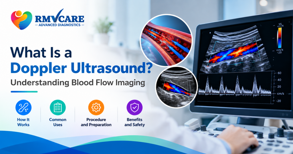
What Is a Doppler Ultrasound? Understanding Blood Flow Imaging
Have you ever wondered how doctors check whether blood is flowing properly through your arteries and veins? A Doppler ultrasound is a specialised imaging test that helps evaluate blood circulation inside the body. It allows doctors to assess the direction and speed of blood flow and identify certain abnormalities affecting blood vessels.
Unlike a conventional ultrasound that mainly produces images of internal structures, Doppler ultrasound provides additional information about moving blood cells. It is commonly used to investigate circulation-related concerns, assess blood vessels and monitor blood flow in selected organs.
Understanding what a Doppler ultrasound is, how it works and when it may be recommended can help patients feel more prepared before undergoing the examination.
What Is a Doppler Ultrasound?
A Doppler ultrasound is a non-invasive diagnostic imaging technique that uses high-frequency sound waves to evaluate blood flow through blood vessels.
The examination works on the Doppler effect, which measures changes in the frequency of sound waves reflected by moving red blood cells. A computer processes these changes to provide information about the direction and speed of blood circulation.
Doctors may recommend Doppler ultrasound to examine arteries and veins in different parts of the body, including the neck, arms, legs and abdomen.
It can help identify blood-flow abnormalities, narrowed blood vessels and certain circulation-related conditions.
How Does a Doppler Ultrasound Work?
A Doppler ultrasound examination uses a handheld device called a transducer. The transducer sends sound waves into the body and receives the echoes reflected by moving blood cells.
The ultrasound system analyses these echoes and converts the information into visual images, graphs or colour-coded displays that help assess blood circulation.
Here is how the examination generally works:
- The patient is positioned comfortably on an examination table.
- A special ultrasound gel is applied to the skin over the area being examined.
- The sonographer moves the transducer across the skin.
- Sound waves interact with moving red blood cells inside the blood vessels.
- The ultrasound machine processes the returning signals to display blood-flow information.
- The images and measurements are reviewed by the radiologist.
The procedure is generally painless and does not use ionising radiation.
What Are the Different Types of Doppler Ultrasound?
Different Doppler techniques provide different information about blood circulation. The appropriate method depends on the area being examined and the medical question.
1. Colour Doppler Ultrasound
Colour Doppler uses colour-coded information to display the direction and relative characteristics of blood flow within blood vessels.
It helps doctors assess circulation patterns and identify certain abnormalities in blood flow.
2. Power Doppler Ultrasound
Power Doppler is particularly sensitive to detecting relatively slow or small-volume blood flow in selected situations.
It can help evaluate blood flow in smaller vessels and certain organs. However, it does not provide the direction of blood flow in the same way as colour Doppler.
3. Spectral Doppler Ultrasound
Spectral Doppler displays blood-flow measurements as graphs over time. It helps assess the speed and pattern of blood circulation through a vessel.
Doctors may use this information to investigate abnormal flow patterns or narrowing in blood vessels.
4. Duplex Doppler Ultrasound
Duplex ultrasound combines conventional ultrasound images with Doppler blood-flow measurements.
It allows doctors to examine both the structure of blood vessels and the movement of blood through them.
Why Do Doctors Recommend a Doppler Ultrasound?
A Doppler ultrasound may be recommended when a doctor needs more information about blood circulation in a particular part of the body.
Some common clinical applications include:
1. Detecting Blood Clots
A venous Doppler ultrasound is commonly used to investigate suspected deep vein thrombosis (DVT), a condition involving blood clots in deep veins, particularly in the legs.
The examination helps assess blood flow and identify findings that may indicate a clot.
2. Evaluating Narrowed or Blocked Arteries
Arterial Doppler examinations help assess blood circulation through arteries. They may be recommended when a doctor suspects narrowing or reduced blood flow.
3. Investigating Leg Swelling and Circulation Problems
Unexplained leg swelling, discomfort or certain circulation-related symptoms may lead a doctor to recommend a venous or arterial Doppler examination.
The type of scan depends on the suspected cause.
4. Assessing Carotid Arteries
A carotid Doppler ultrasound examines the major arteries in the neck that supply blood to the brain.
It may help assess plaque buildup, narrowing and blood-flow abnormalities in these vessels.
5. Evaluating Blood Flow to Certain Organs
Doppler ultrasound may also be used to assess blood circulation in selected organs, including the kidneys and liver, when clinically indicated.
6. Monitoring Blood Flow During Pregnancy
Certain obstetric Doppler examinations help evaluate blood flow between the mother, placenta and developing baby.
These scans are recommended by doctors when additional information about fetal or placental circulation is needed.
Which Body Parts Can Be Examined Using Doppler Ultrasound?
Depending on the doctor’s recommendation and the available examination protocol, Doppler ultrasound can evaluate blood flow in different areas.
| Examination | What it evaluates |
|---|---|
| Venous Doppler | Blood flow through selected veins, commonly in the legs |
| Arterial Doppler | Blood circulation through arteries |
| Carotid Doppler | Blood flow through arteries in the neck |
| Renal Doppler | Blood circulation in the kidneys |
| Hepatic Doppler | Blood flow through selected liver blood vessels |
| Obstetric Doppler | Selected blood-flow patterns involving the pregnancy |
Not every Doppler examination is suitable for every patient. Your doctor will recommend the appropriate scan based on your symptoms and clinical history.
How to Prepare for a Doppler Ultrasound?
Preparation depends on the type of Doppler ultrasound and the body part being examined.
Some examinations require little or no preparation, while others may involve specific instructions.
Before your appointment, consider the following:
- Follow fasting instructions: Certain abdominal or renal Doppler examinations may require fasting. Confirm this with the diagnostic centre.
- Wear comfortable clothing: Loose clothing can make it easier to access the examination area.
- Carry medical records: Bring your prescription, previous scan reports and relevant medical documents.
- Inform the medical team: Mention your medical history, current medications and any previous vascular procedures.
- Follow individual instructions: Do not stop prescribed medication unless your doctor specifically advises you to do so.
Always confirm the preparation requirements for your particular examination before visiting the diagnostic centre.
What Happens During a Doppler Ultrasound Examination?
A Doppler ultrasound is generally performed while the patient is lying down or sitting in a comfortable position, depending on the area being examined.
The sonographer applies ultrasound gel to the skin and gently moves the transducer over the examination area.
The machine captures images and evaluates blood-flow patterns. You may hear soft whooshing sounds from the equipment as it processes the Doppler signals.
The examination is usually painless, although mild pressure from the transducer may occasionally cause temporary discomfort, particularly if the area is already tender.
Once the examination is complete, the gel is removed and the images are reviewed for reporting.
The duration depends on the type of examination and the area being assessed.
What Are the Benefits of Doppler Ultrasound?
Doppler ultrasound is an important diagnostic imaging technique because it provides information about blood circulation without requiring an invasive procedure.
Some of its key benefits include:
- Non-invasive examination: It generally does not require needles or surgical procedures.
- No ionising radiation: It uses sound waves instead of X-rays.
- Real-time blood-flow assessment: It helps doctors evaluate the movement of blood through vessels.
- Evaluation of circulation: It can help investigate certain blockages, narrowing and blood-flow abnormalities.
- Multiple clinical applications: It can examine selected arteries, veins and organ blood vessels.
- Useful for follow-up: It may help monitor blood flow after certain vascular treatments or procedures.
Is Doppler Ultrasound Safe?
Doppler ultrasound is generally considered a safe imaging technique when performed appropriately by trained professionals.
It uses sound waves rather than ionising radiation and is non-invasive in most examinations.
However, ultrasound should still be performed when medically indicated and according to appropriate clinical protocols.
The doctor will determine whether a Doppler examination is suitable based on the patient’s condition and diagnostic requirements.
Doppler Ultrasound vs Regular Ultrasound: What Is the Difference?
Both examinations use high-frequency sound waves, but they provide different types of information.
| Feature | Regular Ultrasound | Doppler Ultrasound |
|---|---|---|
| Main purpose | Produces images of internal structures | Evaluates blood movement |
| Blood-flow assessment | Not its primary function | Yes |
| Technology | Conventional ultrasound imaging | Doppler-based sound-wave analysis |
| Common applications | Abdominal organs, pelvic structures and pregnancy imaging | Arteries, veins and selected organ circulation |
| Radiation | No ionising radiation | No ionising radiation |
In some cases, both techniques are combined during a single examination to provide structural and blood-flow information.
When Should You Ask Your Doctor About a Doppler Ultrasound?
Your doctor may recommend Doppler imaging if you experience symptoms or have medical findings that require assessment of blood circulation.
Examples include:
- Unexplained swelling in one or both legs.
- Leg discomfort or symptoms that may indicate poor circulation.
- Suspected blood clots.
- Certain changes in pulses or blood-flow measurements.
- Follow-up after selected vascular procedures.
Symptoms such as sudden leg swelling, pain, redness or warmth require prompt medical assessment. Sudden breathlessness, chest pain or coughing up blood can indicate a medical emergency and require immediate emergency care rather than waiting for a routine scan.
A Doppler ultrasound should be performed based on medical advice rather than self-diagnosis.
Diagnostic Imaging at RM VCare Advanced Diagnostic Center, Kandivali
Choosing the appropriate diagnostic examination is an important part of the healthcare journey. Understanding the purpose of a scan, its preparation requirements and the information it provides can help patients approach their appointments with greater confidence.
RM VCare Advanced Diagnostic Center is located in Kandivali East, Mumbai, and provides diagnostic imaging services, including 3D/4D sonography and other specialised investigations.
If your doctor has recommended a Doppler ultrasound, contact RM VCare to confirm the availability of the specific examination, appointment requirements and preparation instructions.
For enquiries and appointment information, visit the RM VCare Advanced Diagnostic Center website.
Frequently Asked Questions About Doppler Ultrasound
1. What is a Doppler ultrasound used for?
A Doppler ultrasound evaluates blood flow through arteries and veins. It can help investigate blood clots, narrowed blood vessels and certain circulation-related abnormalities.
2. Is a Doppler ultrasound painful?
No. It is generally painless and non-invasive. A small amount of gel is applied to the skin, and a handheld transducer is moved over the examination area.
3. Does Doppler ultrasound use radiation?
No. Doppler ultrasound uses high-frequency sound waves and does not use ionising radiation like X-rays or CT scans.
4. How long does a Doppler ultrasound take?
The duration varies depending on the body part and type of examination. Your diagnostic centre can provide an estimated appointment duration.
5. Is fasting required before a Doppler ultrasound?
Fasting is not required for every Doppler examination. Certain abdominal or renal Doppler studies may require preparation, so follow the instructions provided by your doctor or diagnostic centre.
6. Can Doppler ultrasound detect blood clots?
Yes. Venous Doppler ultrasound is commonly used to investigate suspected blood clots, particularly deep vein thrombosis in the legs.
7. What is the difference between colour Doppler and normal ultrasound?
A conventional ultrasound mainly produces images of internal structures, while colour Doppler adds colour-coded information about blood-flow direction and relative flow characteristics.
8. Is Doppler ultrasound safe during pregnancy?
Doppler ultrasound may be used during pregnancy when clinically indicated. The examination should be recommended and performed according to appropriate medical protocols.
9. Do I need a doctor’s prescription for a Doppler ultrasound?
The need for a prescription depends on the examination and the diagnostic centre’s requirements. It is best to consult your doctor and confirm the booking requirements beforehand.
10. Where can I enquire about Doppler ultrasound in Kandivali?
You can contact RM VCare Advanced Diagnostic Center in Kandivali East to enquire about the availability of the specific Doppler examination recommended by your doctor.
Conclusion
A Doppler ultrasound is a valuable diagnostic imaging technique that helps doctors evaluate blood circulation through arteries, veins and selected organs. By providing information about the direction and speed of blood flow, it can support the investigation of several vascular and circulation-related conditions.
Understanding how Doppler imaging works, why it may be recommended and how to prepare for the examination can help you make informed decisions about your diagnostic care.
If your doctor has advised a Doppler examination, consult the medical team to confirm the appropriate scan and preparation requirements. For diagnostic service enquiries in Kandivali East, connect with RM VCare Advanced Diagnostic Center.
Medical disclaimer: This article is for general educational purposes only. It does not replace professional medical advice, diagnosis or treatment. Always consult your doctor regarding the appropriate diagnostic examination